A collection of images showcasing advanced microscopy techniques and scientific imaging.
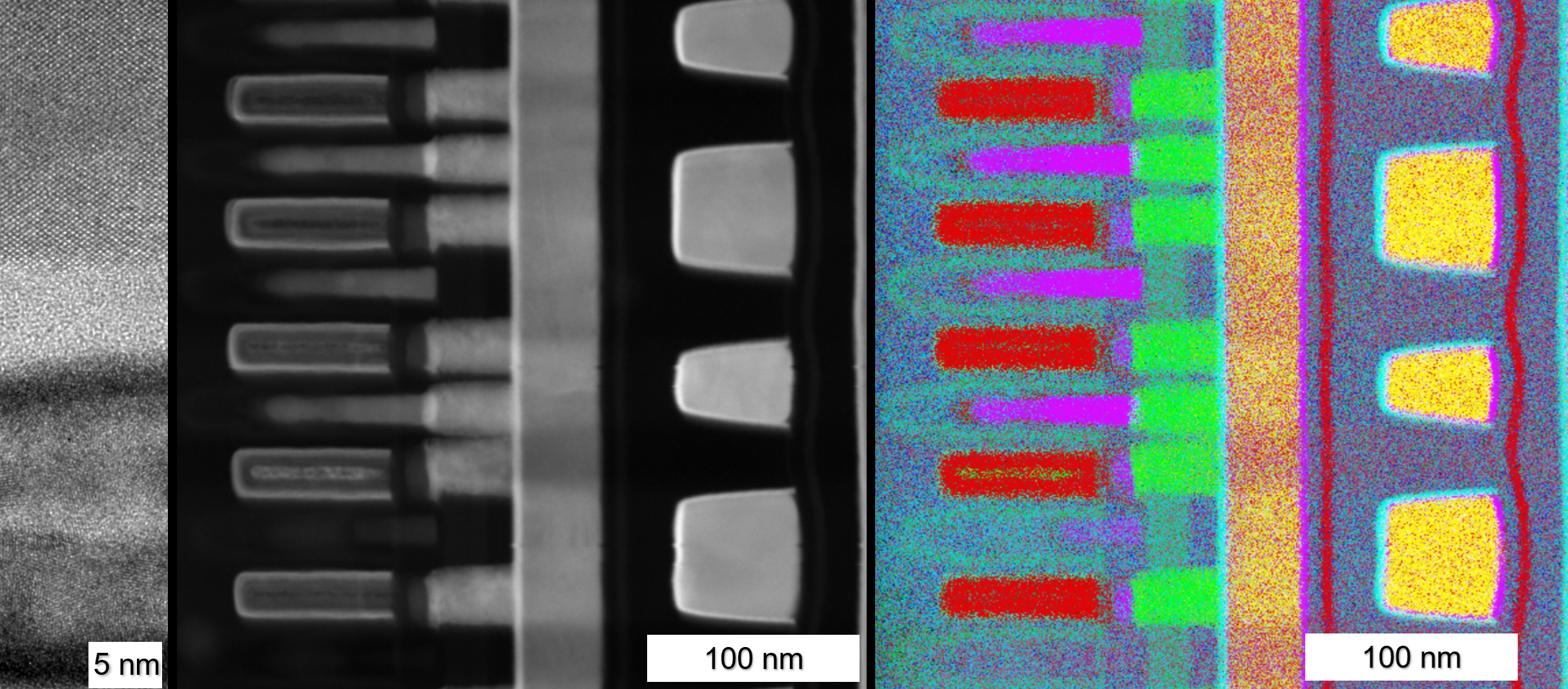
Left) TEM brightfield image of a FinFET including the junction. Middle) STEM HAADF of FinFETs and some interconnects. Right) EDS map of the same. Coloration includes all elements in the map (more than 8) and is generated using an autoencoder neural network (Gainsforth & Dominguez et al., 2024, doi:10.1111/maps.14161)
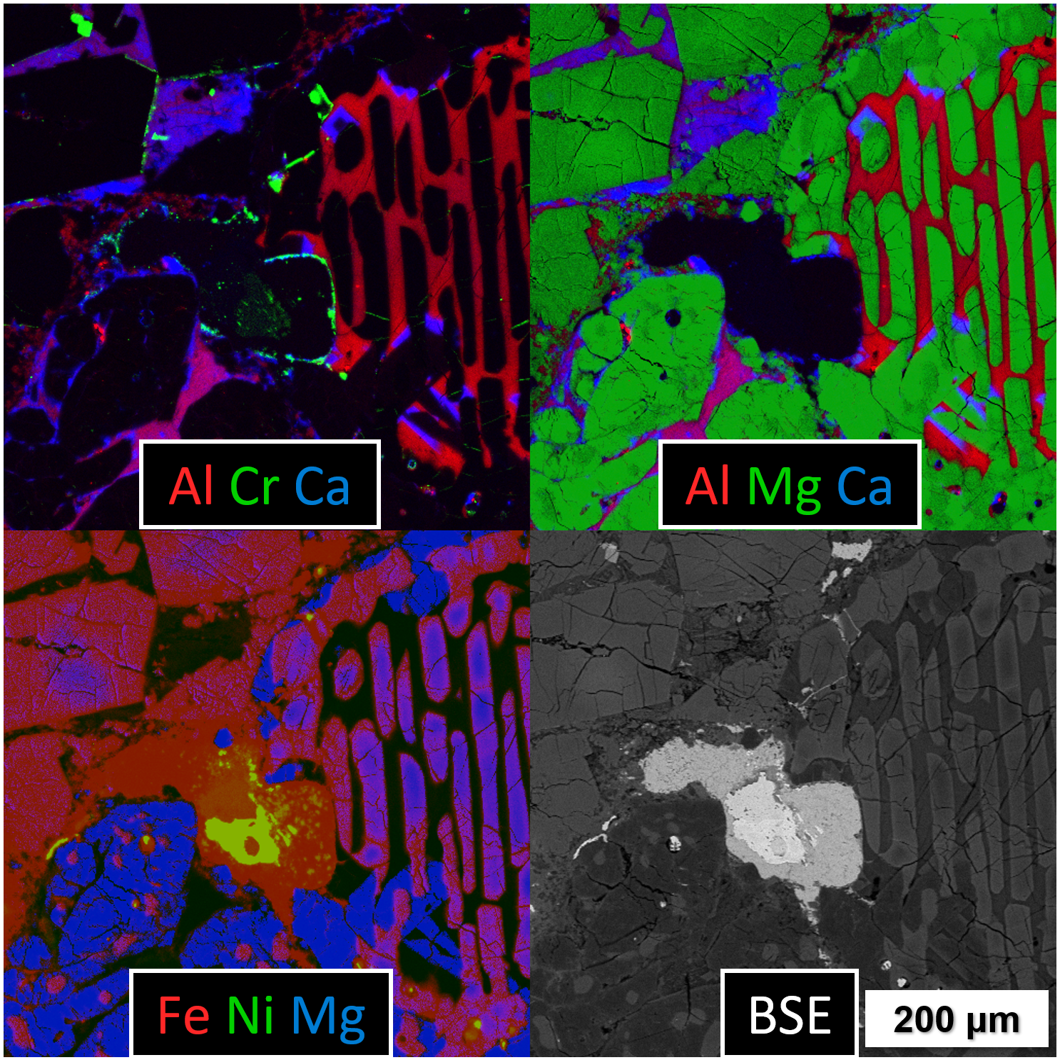
SEM backscatter and EDS images of the Parnallee meteorite showing a unique intersection of a barred and prophyritic chondrule.
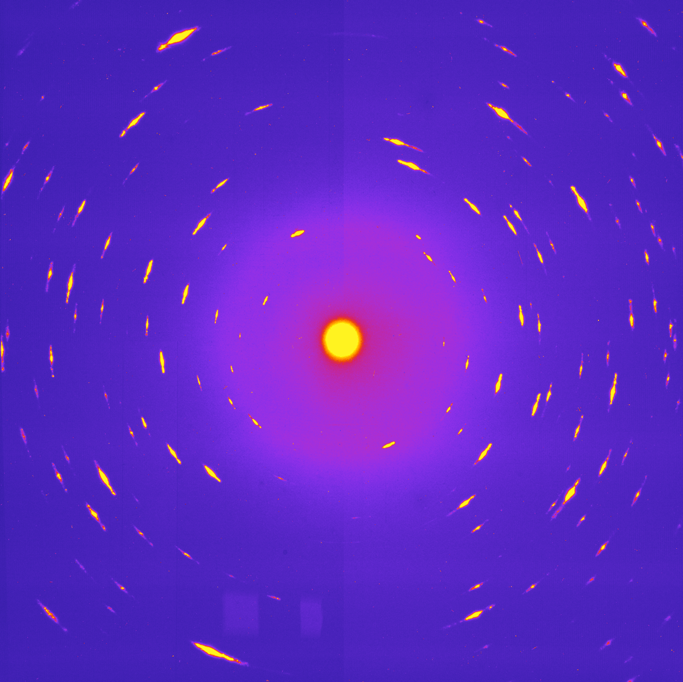
X-ray diffraction pattern of the first verified crystalline interstellar dust candidate obtained from NASA's Stardust mission.
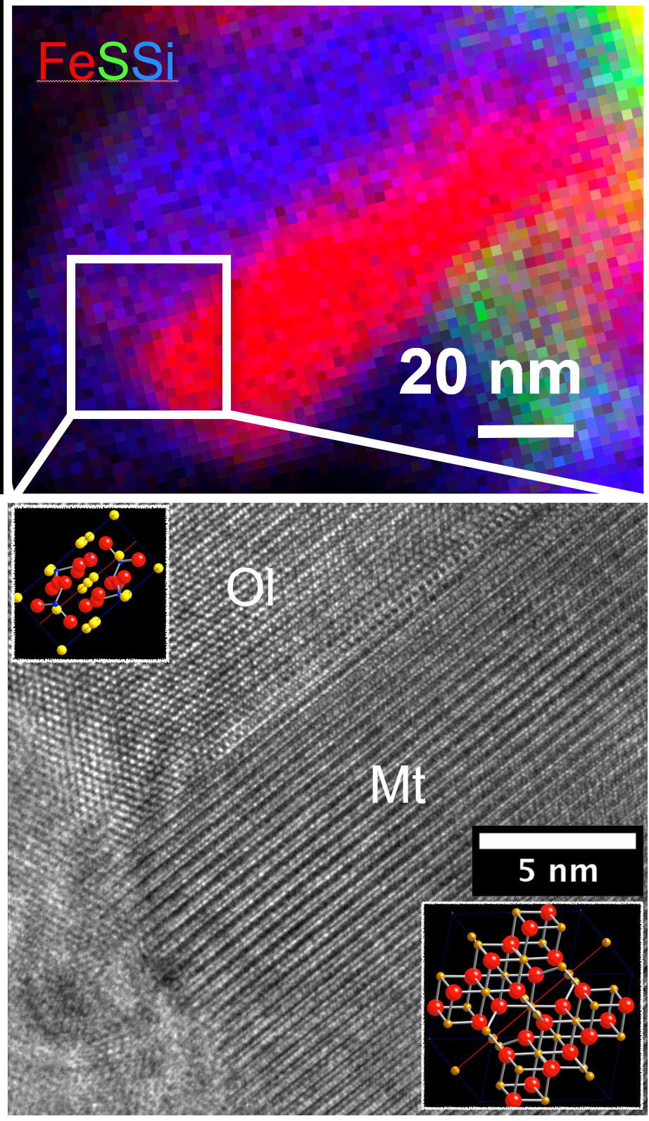
High-resolution Transmission Electron Microscopy (HRTEM) image of a magnetite-olivine interface in an interplanetary dust particle. Additional surrounding silicate is amorphous. This was formed during atmospheric entry as the edge of the dust grain entered the atmosphere at hypersonic velocities. The sample was prepared by FIB.